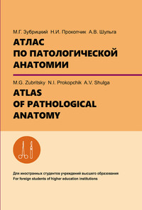
Атлас по патологической анатомии = Atlas of pathological anatomy
Покупка
Новинка
Тематика:
Общая патология. Патофизиология
Издательство:
Вышэйшая школа
Год издания: 2021
Кол-во страниц: 268
Дополнительно
Вид издания:
Учебное пособие
Уровень образования:
ВО - Специалитет
ISBN: 978-985-06-3376-7
Артикул: 821130.01.99
Доступ онлайн
В корзину
Пособие содержит описание макрофотографий учебных препаратов, операционных и секционных наблюдений с необходимыми указателями, подписями теоретическим материалом, включающим современную классификацию, осложнения и исходы. Предназначено для студентов факультета иностранных учащихся с английским языком обучения (специальность 1-79 01 01 «Лечебное дело»). Составлено в соответствии с действующей типовой учебной программой по патологической анатомии, утвержденной Министерством здравоохранения Республики Беларусь.
Скопировать запись
Фрагмент текстового слоя документа размещен для индексирующих роботов.
Для полноценной работы с документом, пожалуйста, перейдите в
ридер.
УДК 616-091(084.42)(075.8)-054.6 ББК 52.5я73 З-91 А в т о р ы : М.Г. Зубрицкий – доцент кафедры патологической анатомии, кандидат медицинских наук; Н.И. Прокопчик – доцент кафедры патологической анатомии, кандидат медицинских наук; А.В. Шульга – доцент кафедры патологической анатомии, кандидат медицинских наук Ре ц е н з е н т ы: кафедра патологической анатомии УО «Белорусский государственный медицинский университет» (доцент кафедры кандидат медицинских наук В.В. Са- вош); доцент кафедры хирургии с курсом патологической анатомии ГУО «Белорусская медицинская академия последипломного образования» кандидат медицинских наук, доцент Ю.И. Рогов; доцент кафедры современных технологий перевода УО «Минский государственный лингвистический университет» кандидат медицинских наук, доцент Т.И. Голикова Все права на данное издание защищены. Воспроизведение всей книги или любой ее части не может быть осуществлено без разрешения издательства и авторов. ISBN 978-985-06-3376-7 Зубрицкий М.Г., Прокопчик Н.И., Шульга А.В., 2021 Оформление. УП «Издательство “Вышэйшая школа”», 2021
ABBREVIATIONS

AIDS – Acquired immunodefi ciency syndrome
AH – Arterial hypertension
ACTH – Adrenocorticotropic hormone
CSF – Cerebrospinal fl uid
COPD – Chronic obstructive pulmonary disease
DIC – Disseminated intravascular coagulation
DNA – Deoxyribonucleic acid
MRI – Magnetic resonance imaging
TNM  – Tumour-node-metastasis
PAP test – Papanicolaou test
PSA – Prostate-specifi c antigen
CHF – Chronic heart failure
CKD – Chronic kidney disease
CLL – Chronic lymphocytic leukemia
COPD – Chronic obstructive pulmonary disease
GH – Growth hormone
CEA – Carcinoembryonic antigen
EMA – Epithelial membrane antigen
IARC – International Agency for Research on Cancer
IHC – Immunohistochemical
PCR – Polymerase chain reaction
RCC – Renal cell carcinoma
SMA – Smooth muscle actin
SCC – Squamous cell carcinoma
hCG – Human chorionic gonadotropin
WHO – World Health Organisation
PREFACE

Pathological Anatomy is an important part of medicine, which provides
the integration of theoretical knowledge with clinical subjects. Th e study of
Pathological Anatomy is divided into General Pathology and Organ System
(Special) Pathology.
General Pathological Anatomy is concerned with basic reactions of cells
and tissues to abnormal stimuli. Th e study is based on independent investiga-
tions of pathologically changed separate organs at macroscopical, histologi-
cal, and electron-microscopical levels.
All illustrations are accompanied by detailed and relevant explanations
with the purpose to facilitate a better understanding of the disease process.
We are convinced that the creation of the present learning tool containing
a wide range of images of macropreparations and museum specimens would
help students to acquire a deep knowledge of pathology and form the basis for
clinical studies.
Th e illustrations are from the teaching collections of several pathology
and university departments. Th e authors are very grateful to A. Plotsky,
A. Grib, V. Aleksinsky for the assistance of the material of this work.
We welcome the opinions of our readers to ensure greater usability in
 future editions (you can contact us by patan@grsmu.by).
INTRODUCTION

Pathology (word derived from the Ancient Greek  pathos
meaning “suff ering” and logy “study of”) is a signifi cant fi eld
in modern medical diagnosis and medical research, concerned
mainly with the causal study of a disease, whether caused by
pathogens or non-infectious physiological disorder.
Pathology consists of abnormalities in normal anatomy
and normal physiology owing to the disease. Pathology
forms a bridge between the initial learning phase of pre-
clinical sciences and the fi nal phase of clinical subjects.
Human pathology is studied under two broad d i v i s i o n s:
• General pathology: deals with general principles of a disease.
• Systemic pathology: that includes the study of a disease pertaining to
the specifi c organ and body systems.
Morphological b r a n c h e s essentially involve the application of the mi-
croscope as an essential tool for the study and include histopathology, cytopa-
thology, and hematology.
Histopathology: it is used synonymously with anatomic pathology, patho-
logical anatomy, morbid anatomy, or tissue pathology and is one of the best
methods. Th e study includes structural changes by naked eye examination
referred to as gross or macroscopic changes and the changes detected by mi-
croscopy, which may be further supported by numerous special staining
methods such as histochemistry and immunohistochemistry to arrive at the
most accurate diagnosis.
Cytopathology: though a branch of anatomic pathology, cytopathology
has developed as a distinct subspeciality in recent times. Cytopathology is
commonly used to investigate diseases involving a wide range of body sites,
oft en to aid in the diagnosis of tumours but also in the diagnosis of some in-
fectious diseases and other infl ammatory conditions. For example, a common
application of cytopathology is the Pap test, a screening tool used to detect
precancerous cervical lesions that may lead to cervical cancer.
It includes the study of cells shed off  from the lesions (exfoliative cytology)
and fi ne-needle aspiration cytology (FNAC) of superfi cial and deep-seated
lesions for diagnosis.
Hematology: It deals with the disease of the blood. Hematology includes
laboratory hematology and clinical hematology; the latter covers the manage-
ment of a patient as well.
Surgical pathology: it deals with the study of the tissue removed from the
living body by biopsy or surgical resection. Surgical pathology forms the bulk
of the tissue material for the pathologist and includes the study of the tissue
by a conventional paraffi  n embedding technique; an incorporative frozen sec-
tion may be employed for rapid diagnosis.
Experimental pathology: this is defi ned as a production of a disease in the
experimental animal and the study of morphological changes in organs aft er
sacrifi cing the animal. However, all the fi ndings of experimental work in ani-
mals may not be applicable to human beings due to specifi c diff erences.
Forensic pathology and autopsy: it includes the study of organs and tis-
sues removed at postmortem for medicolegal work and for determining the
underlying sequence and the cause of death. Th e postmortem anatomical di-
agnosis is helpful to the clinicians to enhance their knowledge about the dis-
ease and their judgment while the forensic autopsy is helpful for the medico-
legal purpose. Th e signifi cance of a careful postmortem examination is ap-
propriately summed up in the old saying: “Th e dead teach the living”.
Non-morphological b r a n c h e s include the following sections:
Clinical pathology: the analysis of various body fl uids such as blood,
urine, semen, CSF, and other fl uids. Th is analysis may be qualitative and
semi-quantitative.
Clinical biochemistry: quantitative determination of various biochemical
constituents in serum and plasma and in the other body fl uids is included in
clinical biochemistry.
Microbiology: it includes the study of disease-causing microbes implicated
in a human disease. Depending upon the type of microorganisms studied, it
has further developed into bacteriology, parasitology, mycology, virology, etc.
Immunology: the detection of the abnormalities in the immune system of
the body comprises immunology and immunopathology.
Medical genetics: the branch of medicine that involves the diagnosis and
management of hereditary disorders.
Molecular pathology: the detection and diagnosis of the abnormalities at
the level of DNA of the cell are now used in diagnostic pathology reports. Mo-
lecular pathology is commonly used in the diagnosis of cancer and infectious
diseases. Techniques are numerous but include a polymerase chain reaction
(PCR), DNA microarray, in situ hybridisation, in situ RNA sequencing, DNA
sequencing, antibody-based immunofl uorescence tissue assays, molecular pro-
fi ling of pathogens, and analysis of bacterial genes for antimicrobial resistance.
Chapter 1

PARENCHYMATOUS, STROMAL-
VASCULAR AND MIXED DYSTROPHIES

1.1 Fatty dystrophy of the liver
(fatty liver disease)

Th e organ has the fl abby consistency, it is increased in the sizes, with
rounded margins, in section – a yellow-brown or ocherous-yellow colour
(the “goose” liver). Causes of the development: hypoxia (in the diseases of the
cardiovascular and respiratory systems); intoxication (alcohol, hepatotropic
poisons); infections (viral hepatitis); endocrine disorders; nutritional (avita-
minosis). Depending upon the cause and amount of accumulation, fatty
change may be mild and reversible or severe producing irreversible cell injury
and cell death. Even a severe form of the fatty liver may be reversible if the
liver is given time to regenerate, and progressive fi brosis has not developed.
Otherwise, the outcome is cirrhosis.
1.2 Hyperkeratosis of the skin (“cutaneous horn”)

Th e surgical material was a fl ap of the skin of the scalp, on which focal
hyperkeratosis like a horn was detected.
“Cutaneous horn” is a rare disease that is diagnosed in people of old age;
somewhat more common in women. Th e place of localisation is predomi-
nantly the area of the face (on the skin of the ears, on the cheeks) and on the
head (its hairy part).
“Cutaneous horn” may occur due to the progression of senile keratosis,
damage to the wart, or papilloma. Th e provoking factors for the formation of
skin horns are: excessive insolation, skin injury, and chronic infl ammation
(viral or other etiology) caused by this background.
Externally, the skin horn is very noticeable and brings a lot of negative
feelings to the patient, though the most dangerous thing is malignancy, but
for the most part, the horns are nothing to worry about.
1

1.3 Ovarian cyst (endometrioid cystadenoma)

Th e ovary is sharply enlarged, deformed, and a plurality of cysts is found
in the incision, fi lled with thick jelly-like contents of a brown colour. Macro-
scopically, endometrioid cystadenoma does not have specifi c features, if the
possible presence of the foci of endometriosis with characteristic features
(“chocolate cysts”) is not taken into account. Endometrioid tumours are epi-
thelial ovarian tumours formed by cells that resemble those of the internal
lining of the uterus (the endometrium). Most oft en, the tumour appears in
women who are between the ages of 20–50. Th e size can reach 15 cm. Th e
histological examination reveals that the lining is represented by layers of
prismatic cells secreting mucus. Th e mucus contains glycoproteins and red
blood cells.
1.4 Amyloidosis of the spleen (“sago spleen”)

Amyloidosis of the spleen oft en causes moderate or even marked enlarge-
ment (200 to 800 gm). Th e deposits may be virtually limited to the splenic
follicles, producing tapioca-like granules on gross examination (“sago spleen”);
the spleen is fi rm in consistency. Th e presence of blood in splenic sinuses usu-
ally imparts a reddish colour to the waxy, friable deposits. Th e cut surface of
the spleen containing white pulp amyloid shows multiple pale foci scattered
throughout the spleen, an appearance labeled sago spleen. Th e amyloid depos-
its begin in the walls of the arterioles of the white pulp and may subsequently
replace the follicles. Staining with Congo Red (CR) is a qualitative method
used for the identifi cation of amyloids in vitro and in tissue sections.
Доступ онлайн
В корзину


