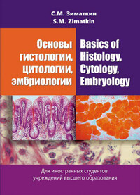
Основы гистологии, цитологии, эмбриологии = Basics of Histology, Cytology, Embryology
Покупка
Новинка
Тематика:
Медицинская биология. Гистология
Издательство:
Вышэйшая школа
Автор:
Зиматкин Сергей Михайлович
Год издания: 2020
Кол-во страниц: 235
Дополнительно
Вид издания:
Учебное пособие
Уровень образования:
ВО - Специалитет
ISBN: 978-985-06-3204-3
Артикул: 821161.01.99
Доступ онлайн
В корзину
В пособии коротко и доступно изложены основные вопросы цитологии (учение о клетке), общей (учение о тканях) и частной гистологии (микроскопическая организация органов) и эмбриологии (развитие зародыша и плода)
человека. Для студентов учреждений высшего образования, обучающихся на английском языке.
The basic issues of cytology (cell biology), general (structure of tissues) and particular histology (microscopic organization of organs), and embryology (embryo development) are shortly and simply described in the present manual. The book is dedicated to students of medical higher education institutions studying in English.
Тематика:
ББК:
УДК:
ОКСО:
- ВО - Бакалавриат
- 45.03.02: Лингвистика
- ВО - Специалитет
- 30.05.01: Медицинская биохимия
- 30.05.02: Медицинская биофизика
- 31.05.01: Лечебное дело
- 32.05.01: Медико-профилактическое дело
ГРНТИ:
Скопировать запись
Фрагмент текстового слоя документа размещен для индексирующих роботов.
Для полноценной работы с документом, пожалуйста, перейдите в
ридер.
                С.М. Зиматкин


Основы Basics of гистологии, Histology, цитологии, Cytology, эмбриологии Embryology


Допущено
Министерством образования Республики Беларусь в качестве учебного пособия для иностранных студентов учреждений высшего образования по специальности «Лечебное дело»


Минск «Вышэйшая школа»
2020
УДК [611.018+611.013](075.8)-054.6
ББК 28.70я73

     З-62


   Рецензенты: кафедра гистологии, цитологии и эмбриологии УО «Белорусский государственный медицинский университет» (заведующий кафедрой кандидат медицинских наук, доцент Т.М. Студеникина); кафедра гистологии, цитологии и эмбриологии УО «Витебский государственный медицинский университет» (доцент кафедры кандидат биологических наук, доцент В.Н. Грушин; заведующий кафедрой доктор медицинских наук, профессор О.Д. Мяделец); доцент кафедры современных технологий перевода УО «Минский государственный лингвистический университет» кандидат филологических наук, доцент Т.И. Голикова


    Все права на данное издание защищены. Воспроизведение всей книги или любой ее части не может быть осуществлено без разрешения издательства.


ISBN 978-985-06-3204-3

                    © Зиматкин С.М., 2020
                    © Оформление. УП «Издательство

“Вышэйшая школа”», 2020
FOREWORD


    The present book is a short course of modern medical histology, cytology, and embryology. The author set the goal to make it interesting and fascinating, concise, simple and understandable, devoiding of minor details, but still containing the necessary minimum of knowledge for medical students. The manual should help them understand the microscopic structure and organization of tissues and organs of the human body, as well as the functioning principles of their constituent cells and tissues (cyto-and histophysiology).
    The text is as structured as possible (divided into paragraphs, terms are highlighted in bold type and/or given italics, emphatic points are given with increased letter-spacing), resulting in easier visual perception. Less important (in the author’s opinion) information is given in a smaller font. All terminology is brought into line with the modern international histological nomenclature.
    The manual is elaborated following the current standard curriculum in Histology, Cytology, and Embryology designed for students of medical higher education institutions studying in English.
    We hope that it will help medical students to study and understand this difficult, thus interesting and essential subject for future doctors, allowing them to put learning into practice.
    This book can partially replace voluminous, complex and expensive international textbooks on this subject, which do not fully correspond to the program of the Belarusian medical institutions and are difficult for most students to understand.
    The author expresses gratitude to T.V. Klimut, the laboratory assistant of our (the Grodno State Medical University) Department of Histology, Cytology, and Embryology for her technical assistance in preparing the book script for publication.


Professor S.M. Zimatkin
                1.  INTRODUCTION TO HISTOLOGY


   The word “histology” (derived from the Greek histos — “tissue” and logos — “study” or “science”) means the science of tissues.
   Our course consists of 4 parts:
   1. General Histology — the science about the tissues.
   2.    Particular Histology — the study of the microscopic structure of organs (microscopic anatomy).
   3. Cytology — cell biology.
   4. Embryology — the study of embryo development.
   Therefore, the full name of the given course is “Histology, Cytology, Embryology”.

        Units of length used in Histology

   •     1 micrometer (mcm, ц) — 10⁻⁶ m (used in the light microscopy for the measurements of cells);
   •     1 nanometer (nm) — 10⁻⁹ m (used in the electron microscopy for the measurement of subcellular structures).
   Therefore, the main methods used in histology are microscopic and the main instruments for studies are microscopes. To study tissues and organs by microscopes it is necessary to make the histological preparations of them.

        Making histological preparations for a light microscopy

   To avoid tissue digestion by enzymes (autolysis) or bacteria and to preserve its initial structure, the pieces of organs should be promptly and adequately fixed after removal from the body. The chemical substances used to preserve tissues are called fixatives and there are hundreds of them. One of the best and most popular fixatives for routine light microscopy is a 4% solution of formaldehyde; the mixture of ethanol, chloroform, and acetic acid are also widely used.
   After that, the samples should be dehydrated (in the battery of alcohols of increasing concentration, usually from 70% to 100% ethanol) and cleared (with organic solvents like xylene). Once the tissue is impregnated with the solvent, it is placed in melted paraffin in the oven, usually at 58-60 °C.

4
   Since tissues and organs embedded in paraffin are usually too thick for transillumination in a light microscope, they must be cut to obtain thin, translucent sections by fine cutting instruments called microtomes. The small paraffin blocks containing the tissues are sectioned by the microtome’s steel knife to a thickness of 1—10ц. Then the sections are transferred to a histological glass for staining (Fig. 1.1).


Fig. 1.1. Making paraffin sections (by E. Ulumbecov, Yu. Chelishev)

   To see the tissue structures under the microscope it is necessary to specifically stain them.
   Types of dyes and staining:
   •     basophilia (from the Greek fileo — “love”, i.e. loving the basic dyes) — basic (hematoxylin) — the ability to be stained by basic dyes;
   •     oxyphilia (“loving the acid dyes”) — acid (eosin) — the ability to be stained by acid dyes;
   •     polychromatophilia — the ability to be stained by both types of dyes;
   •     metachromasia — the ability of structures to change the color of dye (e.g. the blue dye stains the structures in red).
   Of all dyes, the combination of hematoxylin and eosin (H&E) is the most commonly used. Hematoxylin stains the cell nucleus in blue. In contrast, eosin stains the cytoplasm in red. Many other dyes are used in different histological procedures.

5
   In addition to tissue staining with dyes, impregnation with metals (as silver) is a common method, especially in studies of the nervous system.

        Microscopes

   The microscope is composed of the following parts:
   •     mechanical;
   •     optical.
   The optical components consist of three systems of lenses:
   •     condenser;
   •     objective;
   •     ocular.
   The condenser collects and focuses the light that illuminates the object to be observed.
   The objective and ocular lens enlarges and projects the illuminated image of the object in the direction of the viewer’s eye retina. The total magnification is obtained by multiplying the magnifying power of the objective and ocular lenses.
   Resolution (resolving power) — the smallest distance between two points at which they can be seen as separate objects.

Light microscopy

   With the light microscope, stained preparations are usually examined through transillumination.
   The maximal resolution of the light microscope is approximately 0.2 ц. Objects smaller than 0.2 ц cannot be distinguished with that microscope. The quality of the image — its clarity and richness of details — depends on the microscope’s resolution. It depends mainly on the quality of its objective lens.

   Conventional light, phase contrast, polarizing, confocal, and fluorescence microscopy are all based on the interaction of photons and tissue components.

   There are some problems in the interpretation of histological preparations. There is a tendency to think in terms of only two dimensions when examining thin sections, while the structures from which the sections are made, actually have three dimensions.
   Another difficulty in the study of microscopic preparations is the impossibility of differentially staining all tissue components on only one slide. It is, therefore, necessary to examine several preparations stained by


6
different methods before a general idea of the composition for the structure of any type of tissue to be obtained.


Electron microscopy

   Electron microscopy is based on the interaction of the beam of electrons and tissue components. The electron microscope is an imaging system that permits high resolution (0.1 nm). This by itself permits enlargements to be obtained up to 1000 times greater than those obtained with light microscopes. The image obtained is finally seen on a fluorescent screen.
   Transmission electron microscopy requires a much thinner section (0.02—0.1 ц) of the samples embedded in hard epoxy plastic. The blocks thus obtained are so hard that the glass or the diamond knives are usually necessary to section them. Since the electron beam in the microscope cannot penetrate glass, the extremely thin sections are collected on small metal grids. Those portions of the section spanning the holes in the mesh of the grid can be examined in the microscope.
   Scanning electron microscopy permits three-dimensional views of the surfaces of cells, tissues, and organs. The very narrow (10 nm) electron beam is moved sequentially from point to point across the surface of the sample. At each point, the primary electron beam interacts with a thin metal coating previously applied to the specimen and produces reflected or emitted electrons. The electron signal fluctuation is captured by a detector.

        Histochemistry

   These methods are used mainly to localizing different substances and activities of enzymes in tissue sections. The methods usually produce insoluble colored or electron-dense compounds that enable the localization of specific substances through light or electron microscopy.
   DNA can be identified and quantified in cell nuclei using the Feulgen reaction, which produces a red color in the presence of DNA.
   Glycogen can be demonstrated by the periodic acid-Schiff (PAS) reaction producing a new complex compound with a purple color.
   The dyes most commonly used for l ipids are:
   •    Sudan IV;
   •    Sudan black.
   These dyes confer red and black colors, respectively.

7
   In its turn, most enzymatic (enzymes) histochemical procedures are based on the production of intensely stained or electron-dense precipitates at the site of enzymatic activity.

        Immunocytochemistry

   When a tissue section containing certain antigens is incubated in a solution containing labeled antibodies to these antigens, the antibodies bind specifically to the antigens, the location of which can then be seen with either the light or an electron microscope.

        Hybridization techniques

   It is based on the ability of previously labeled single-stranded segments of DNA or RNA (probes) to bind specifically to complementary nucleic acids. When applied directly to cells and tissues, hybridization is called in situ hybridization. Through it, one can localize specific DNA sequences (such as genes) or RNA.

        History of histology

   1.    Before the microscopes appeared (started > 2000 years ago). Aristotle — 4th century B.C.; Galen — 3rd century A.D.; Avicenna — 10th century, Vesalius — 16th century.
   2.   Microscopic period (started > 350 years ago).
   •     Inventors of the first microscopes: Galileo Galilei — 1600; the Yancens (father and son) —1610; Cornelius Drebel — 1619;
   •     Robert Hooke analyzed the cellular structure of oak bark — 1665, the book “Micrography”;
   •    Antonie van Leeuwenhoek — discovered protozoa in the water drop;
   •    18th—19th centuries — achromatic microscopes were developed;
   •     Matthias Schleiden and Theodor Schwann formulated the Cell Theory — 1838;
   •     The 60s of 19th century — the first Departments of Histology in Moscow (A.I. Babukhin) and St. Petersburg (F.V. Ovsyannikov) Universities, Russia.
   3.    Modern period (since the middle of the 20th century). It started after the electron microscope creation and the molecular biology methods development.


8
        Cell theory (1838)

   •  The cell is the smallest part of a living organism;
   •  Cells of different organisms have a similar structure;
   •  Every cell derived from another cell (every cell from a cell);
   •     Organisms are the complex ensembles of cells and their derivatives, united into the systems of tissues and organs.


                2.   BASIC CYTOLOGY.
                BIOLOGICAL MEMBRANES.
                CYTOPLASM


   The cell is the smallest alive part of the human body, consisting of cytoplasm and nucleus. It provides the bases of the structure, the development and the functions of the body, but it is dependent on its regulatory mechanisms.
   The cell possesses all five signs of an alive structure:
   1. Definite structural organization;
   2. Metabolism (exchange of substances with the environment);
   3. Constant self-renovation and reproduction;
   4. Irritability and excitability;
   5. Movement.
   There are about 10¹⁴ cells in the human body, divided into 200 types.
   The shapeofcells varies significantly (flat, cuboidal, columnar, round, spindle-like, stellate, etc). The size of the cells is between 5—150 mcm (Fig. 2.1).
   Derivatives of cells:
   •  Postcellular structures (erythrocytes, platelets);
   •     Supracellular structures (symplasts — a lot of cytoplasm and many nuclei);
   •  Intercellular matrix (ground substance and fibers).
   The typical cell of the human body consists of a nucleus and cytoplasm. It is separated from the environment by a cell membrane (plasma membrane, plasmalemma). The cytoplasm contains organelles (the constant, permanent structures of the cells) and inclusions (the temporary structures). The organelles and inclusions are suspended in the cytosol.

9
Lysosomes

Pinocytotic vesicles

Cell center (centrioles)

Ribosomes

Mitochondrion


                                                           ___ Rough endoplasmic reticulum
                                     ----Nucleus
                                     -------Nuclear envelope
                                                          ■—Nucleolus


Secretory vesicles


Golgi complex

Fig. 2.1. Animal cell structure (by V. Eliseev, Yu. Afanasyev, E. Kotovsky)


        UNIVERSAL BIOLOGICAL MEMBRANE


   The cell is divided into compartments by biologic membranes. All of them have quite similar structures and properties which are summarized in the term of the universal biological membrane (Fig. 2.2).


Fig. 2.2. Structure of universal biological membrane (a) and cell membrane (b) (by O. Volkova, O. Eletsky):
1 — phospholipid molecules; 2 — bilayer of phospholipids; 3 — integral (transmembrane) protein; 4 — semi-integral protein; 5 — peripheral proteins; 6 — glycocalyx; 7 — submembrane layer; 8 — microfilaments; 9 — microtubules; 10 — intermediate filament; 11 — glycolipid molecule

10
Доступ онлайн
В корзину


